Description
Accurate and timely diagnosis of malaria is essential for disease management and surveillance. Thin and thick blood smear microscopy and malaria rapid diagnostic tests (RDTs) are standard malaria diagnostics, but both methods have limitations. The novel automated hematology analyzer XN-30 provides standard complete blood counts (CBC) as well as quantification of malaria parasitemia at the price of a CBC. This study assessed the accuracy of XN-30 for malaria detection in a controlled human malaria infection (CHMI) study and a phase 3 diagnostic accuracy study in Burkina Faso.
Methods
Sixteen healthy, malaria-naive CHMI participants were challenged with five Plasmodium falciparum-infected mosquitoes. Blood was sampled daily for XN-30, blood smear microscopy, and malaria qPCR. The accuracy study included patients aged > 3 months presenting with acute febrile illness. XN-30, microscopy, and rapid diagnostic tests (HRP-2/pLDH) were performed on site; qPCR was done in retrospect. The malaria reference standard was microscopy, and results were corrected for sub-microscopic cases.
Results
All CHMI participants became parasitemic by qPCR and XN-30 with a strong correlation for parasite density (R2 = 0.91; p < .0001). The XN-30 accurately monitored treatment and allowed detection of recrudescence. Out of 908 patients in the accuracy study, 241 had microscopic malaria (density 24–491,802 parasites/μL). The sensitivity and specificity of XN-30 compared to microscopy were 98.7% and 99.4% (PPV = 98.7%, NPV = 99.4%). Results were corrected for qPCR-confirmed sub-microscopic cases. Three microscopy-confirmed cases were not detected by XN-30. However, XN-30 detected 19/134 (14.2%) qPCR-confirmed cases missed by microscopy. Among qPCR-confirmed cases, XN-30 had a higher sensitivity (70.9% versus 66.4%; p = .0009) and similar specificity (99.6% versus 100%; p = .5) as microscopy. The accuracy of XN-30 for microscopic malaria was equal to or higher than HRP-2 and pLDH RDTs, respectively.
Conclusions
The XN-30 is a novel, automated hematology analyzer that combines standard hemocytometry with rapid, objective, and robust malaria detection and quantification, ensuring prompt treatment of malaria and malaria anemia and follow-up of treatment response.
Trial registration
Both trials were registered on clinicaltrials.gov with respective identifiers NCT02836002 (CHMI trial) and NCT02669823 (diagnostic accuracy study).
Electronic supplementary material
The online version of this article (10.1186/s12916-019-1334-5) contains supplementary material, which is available to authorized users.
Keywords: Malaria, Diagnosis, Sensitivity, Specificity, Burkina Faso
Background
Malaria remains a major cause of morbidity and mortality around the world. Sub-Saharan Africa is most affected with Plasmodium falciparum accounting for 99% of estimated cases [1]. Timely and accurate diagnosis of malaria is essential for disease management and control [2]. Thin and thick blood smear microscopy (further referred to as microscopy) and malaria rapid diagnostic tests (RDTs) are standard malaria diagnostics in endemic areas. RDTs have significantly improved the use of diagnostics for malaria diagnosis, accounting for 74% of diagnostic tests performed among suspected cases in 2015 [3]. Both techniques provide challenges in clinical practice: RDTs are antigen-directed and relative to their design, can neither quantify malaria parasitemia, nor allow monitoring of treatment response, prerequisites to manage severe malaria. Additionally, RDTs cannot distinguish current from recently treated infections [4]. Consequently, false-positive results may be misinterpreted as treatment failure [5]. WHO therefore reserves a role for microscopy, which is however labor-intensive and its quality is heavily observer-dependent. Molecular tests are complex to perform, require highly trained personnel, and are relatively expensive, limiting its general use.
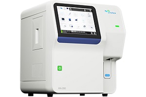
The XN-30 (Sysmex, Kobe, Japan) is a novel automated hematology analyzer and malaria diagnostic that directly detects and quantifies Plasmodium parasites (falciparum and non-falciparum) in blood using violet laser technology [6–8]. Our study aimed to evaluate the accuracy of the XN-30 for the detection of Plasmodium falciparum malaria. First, we assessed its performance in healthy participants in whom low-density parasitemia was induced in a controlled human malaria infection (CHMI) study. To establish the performance under field conditions, we then performed a phase 3 diagnostic accuracy study among febrile patients in Burkina Faso.
Methods
Controlled human malaria infection (CHMI)
The ability of the XN-30 to detect low-density sexual and asexual parasitemia and monitoring of treatment was studied in a CHMI trial (ClinicalTrials.gov, NCT02836002). Details and results of the primary objectives of this study have recently been published [9]. Ethylenediaminetetraacetic acid (EDTA)-anticoagulated venous blood was collected twice daily for XN-30, and results were compared with qPCR for asexual- and qRT-PCR for sexual P. falciparum parasites. Both techniques are described in detail elsewhere [9].
Diagnostic accuracy study
This prospective, double-blinded, phase 3 diagnostic accuracy study (ClinicalTrials.gov, NCT02669823) was performed at the Clinical Research Unit of Nanoro (CRUN) [10]. The primary objectives were (i) to assess the diagnostic sensitivity and specificity of the XN-30 to detect malaria parasitemia in children and adults with an acute febrile illness against thick blood smear microscopy or qPCR in case of a negative thick smear or incongruent results of XN-30 and microscopy and (ii) to assess the diagnostic accuracy of the Infection Manager System (IMS) for the detection of bacterial bloodstream infection in a malaria-endemic area. The secondary objective was to compare the accuracy of the XN-30 analyzer to diagnose malaria compared to malaria RDT. The results of the IMS are reported elsewhere to increase legibility.
Procedures
Nanoro is a rural area of Burkina Faso which is hyperendemic for Plasmodium falciparum, though Plasmodium ovale and Plasmodium malariae are sporadically found [11]. Participants were enrolled between March 2016 and June 2017 at the “Centre Medicale avec Antenne Chirurgicale” (CMA) of Nanoro, to which CRUN is affiliated. Consecutive patients of 3 months and older suspected of acute febrile illness were screened for eligibility. Patients were eligible if they had a measured temperature of ≥ 38.0 °C or ≤ 35.5 °C, or a reported history of fever up to 48 h prior to presentation, or suspicion of severe infection with signs of severe clinical illness. Patients with fever lasting more than 7 days were excluded. Upon inclusion, 2–5 mL EDTA anti-coagulated blood was obtained and analyzed within 1 h after sampling. Laboratory analyses were performed and interpreted by experienced laboratory technicians who were blinded to clinical data. Patients were followed daily during hospitalization, and follow-up samples were taken if clinically indicated. Another follow-up sample was taken at approximately 2 weeks after inclusion.
Data was collected on case report forms (CRFs) and entered into a secure database (RedCap, Vanderbilt University, Nashville, USA) after conformity check by a medical doctor. Entered data were checked against the CRFs by a data manager. Approximately 10 % of participant study files were checked by an independent monitor. Results from qPCR were entered into an Excel (Microsoft, Washington, USA) database and merged with the principal database. The researchers were blinded to the XN-30 until the clinical database was locked. Interpretation of the index test was done by blinded researchers after inclusion was completed. Laboratory procedures were standardized; quality controls were performed according to good clinical and laboratory practices (GCLP) guidelines.
Index test: XN-30
The XN-30 is an automated hematology analyzer (Sysmex Corporation, Kobe, Japan) which can be used for malaria detection as described in detail elsewhere [6]. The analyzer aspirates and dilutes blood samples in a diluent solution (CELLPACK DCL). Subsequently, the nucleic acids are stained with a staining solution (Fluorocell M) along with a lysis solution (LysercellM). Infected red blood cells (iRBC) and white blood cells (WBC) are detected by a violet semiconductor 405 nm laser beam. Parasitemia percentage is calculated by the ratio of infected- and uninfected red blood cells. Output data separately reports a complete blood count (CBC), the presence of gametocytes and parasitemia—both as a percentage of infected red blood cells (MI-RBC%) and absolute parasite density (MI-RBC#) expressed as parasites/μL. The data is automatically determined by analyzing scatter grams plotted three dimensionally with forward-side scattered light, side scattered light, and side fluorescent light. iRBC are visible as a separate RBC population on the scattergram (Fig. 1). In case of abnormal scatter gram distribution, the malaria result is reported as “inconclusive.”
Reviews
There are no reviews yet.