Description
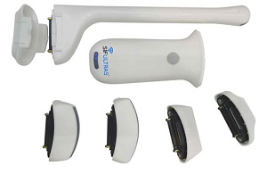
The SIFULTRAS-8.53 Color Doppler Wireless Ultrasound Scanner delivers high-quality imaging with ultimate adaptability. This advanced system is built around a modular probe design with five interchangeable transducer heads, allowing clinicians to swap specialized probes for any clinical scenario. This versatility makes it a powerful, all-in-one, and cost-effective tool for a wide range of medical specialties.
Unlock Unprecedented Versatility: The 5-Head Interchangeable System of the SIFULTRAS-8.53
For healthcare professionals, true diagnostic power lies in adaptability. The SIFULTRAS-8.53 redefines portable ultrasound by offering a comprehensive 5-head interchangeable probe system in one compact, wireless device. Move beyond the limitations of single or dual-head scanners and instantly adapt to any clinical scenario without investing in multiple, cumbersome machines.
This revolutionary approach means:
Unmatched Versatility: Seamlessly transition from deep abdominal scans to high-resolution superficial imaging, cardiac assessments, and specialized transvaginal exams with a simple probe swap.
Superior Cost Efficiency: One scanner delivers the functionality of an entire imaging suite, providing significant savings on capital equipment without compromising diagnostic capability.
Enhanced Clinical Workflow: Streamline examinations and improve patient throughput with quick, intuitive probe changes for the most comprehensive assessments.
Portability with Power: Carry a complete diagnostic imaging lab in your pocket, ready for any scenario from the clinic and bedside to remote point-of-care settings.
The Complete 5-Probe Imaging Suite
1. High-Resolution Convex Probe: For Deep-Penetration Imaging
The convex probe (3.2-4 MHz) is your go-to for in-depth examinations, penetrating from 90 mm to 305 mm to visualize internal organs with clarity.
Primary Applications:
Abdominal (Liver, Kidneys, Gallbladder)
Obstetrics & Gynecology (OB/GYN)
Pelvic and Urology
Cardiac and Lung (Thoracic)
2. High-Frequency Linear Probe: For Superficial Detail & Precision
The linear probe (7.5-10 MHz) offers exceptional resolution for structures close to the surface, with a penetration depth of 40-100 mm.
Primary Applications:
Musculoskeletal (MSK), Nerve, and Small Parts
Vascular Imaging and Thyroid
Breast Examinations
Superficial Lesions and Guided Procedures
3. Phased Array Probe: For Cardiac & Thoracic Imaging
The phased array probe is optimized for dynamic cardiac imaging, providing a small footprint to fit between ribs for clear views of the heart and thoracic organs.
Primary Applications:
Cardiac (Echocardiography)
Thoracic (Lung)
Abdominal (for specific deep, sector-shaped views)
4. Mini Linear Probe: For Ultra-High Resolution & Small Parts
The compact footprint of the mini linear probe provides superior resolution for the most superficial structures and difficult-to-image areas.
Primary Applications:
Vascular Access
Pediatric Imaging
Superficial Tendons and Nerves
Ophthalmic and Testicular Scans
5. Transvaginal Probe: For Specialized Gynecological Diagnostics
The curved array transvaginal probe is designed for high-resolution endocavity imaging, offering detailed visualization of pelvic anatomy.
Primary Applications:
OB/GYN and Infertility
Early Pregnancy
Key Features of the SIFULTRAS-8.53 System
5-Head Interchangeable Design: Convex, Linear, Phased Array, Mini Linear, and Transvaginal probes create a complete wireless imaging suite.
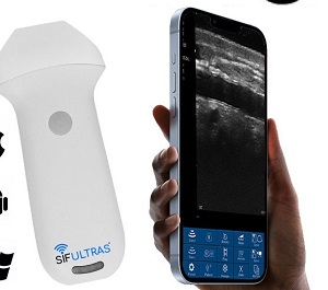
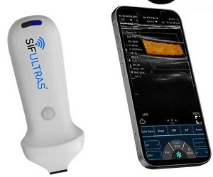
Wireless Connectivity: Stream high-quality color images via WiFi to your iOS or Android phone or tablet.
AI-Enhanced Imaging: Incorporates artificial intelligence and specialized presets to automatically optimize image quality for a smooth, efficient workflow.
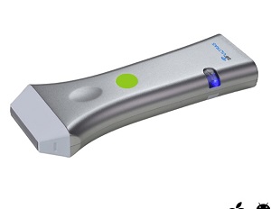
Exceptional Portability: Lightweight and compact for easy use in clinics, hospitals, surgery, and mobile care settings.
Simplified Sterilization: Use with disposable protection covers to easily address infection control protocols.
Ideal for Multi-Specialty Clinical Practice
The SIFULTRAS-8.53 is the preferred choice for medical professionals seeking top-tier, versatile imaging. Its broad application range covers:
Abdomen & Thoracic
Cardiac (with Phased Array)
OB/GYN & Urology (with dedicated Transvaginal probe)
Vascular & Small Parts (with Mini Linear)
MSK, Nerve, & Point-of-Care (POCUS)
Unlocking Blood Flow Dynamics: The Essential Role of Color Doppler Mode
Color Doppler Mode is a vital ultrasound feature that provides a real-time, color-coded map of blood flow velocity and direction directly overlaid on the grayscale anatomical image. It transforms vascular and cardiac assessments from guesswork into precise visualization.
Core Functions & Diagnostic Applications:
Visualize Blood Flow: Get an immediate visual overview of flow patterns within blood vessels and the heart.
Quickly Identify Structures: Rapidly distinguish between blood vessels, cysts, and other structures.
Assess Flow Direction and Velocity: Instantly evaluate hemodynamics with a standard color map (red for flow toward the probe, blue for away).
Locate Blockages and Clots: Pinpoint vessel narrowing (stenosis) or thrombosis by showing disrupted or absent flow.
Detect Vascular Disease: Visualize turbulent flow caused by arterial plaques.
Evaluate Organ Perfusion: Observe blood flow to major organs like the heart, kidneys, and liver.
Guide Precise Measurements: Accurately place the sample volume for Pulsed-Wave Doppler quantification.
Specifications:
• Scan mode: Electronic Array, Electronic Convex & Linear array scanning
• Frequency: Convex 3.2/5.0MHz
Phased array 3.2/4.0MHz,
Linear probe 7.5/10MHz
Mini Linear 10/13.2MHz
Transvaginal 6.6/8.0 MHz
• Scanning Depth: Convex 90/160/220/305mm, Linear 20/40/60/100mm, Transvaginal 20/40/60/100, adjustable
• Field of view: Convex R60, Phasedarray R80, Linear L40
• Elements: 128 elements
• Channels: 32 channels
• Screen: iOS/Android/Windows screen
• Supporting system: iOS, Android, Windows
• Display mode: B, B/M, Color, PDI, PW
Linear array COLOR mode
Deflection angle -12 -7 0 7 12 degrees
PRF ≥ 4 groups
Wall filter 0 – 15
Color gain 0 – 10
Linear array PW mode
Sampling volume: 1, 2, 5 mm
Baseline: 0 – 500 cm/s
PRF ≥ 4 groups
Color gain: 0 – 100
Correction angle: -85 – 85 degrees
Convex Array COLOR Mode
Deflection Angle: Not Available
PRF: ≥ 4 groups
Wall Filter: 0 – 15
Color Gain: 0 – 10
Convex array PW mode
Sampling volume: 1, 2, 5 mm
Baseline: 0 – 800 cm/s
PRF: ≥ 4 groups
Correction angle: -85 to 85 degrees
Cardiac COLOR mode
Deflection angle: Not available
PRF ≥ 4 groups
Wall filter: 0 – 15
Color gain: 0 – 100
Cardiac PW mode
Sampling volume: 1, 2, 5 mm
Baseline: 0 – 600 cm/s
PRF ≥ 4 groups, Note: For USB phased array probe, PRF is 2 – 20K ≥ 8 groups
Correction angle: -85 – 85 degrees
• Image Adjustment: gain, focus, reverse pulse harmonics, noise reduction
• Puncture Auxiliary Function: In-Plane puncture guide, Out-Plane puncture guide, automatic vascular measurement function
• Measure: Thyroid, small organs, pediatrics, blood vessels, carotid artery, breast, musculoskeletal, blood flow, nerves, abdomen,
gynecology, obstetrics, heart, urinary system, kidneys, lungs
• Power: by built-in lithium battery, 2800mAh, wireless charging can extend the working time indefinitely
• Playback : manual and automatic, playback frames to be 100/200/500/1000
• Image gray scale: 256 level
• Image frame rate: 18 frames/second
• Dynamic Range: 40/50/60/70/80/90/100/110
• Image/Video Storage: jpg, png mp4 and DCM
• WiFi Type : 802.11n/2.4G/5G dual-band 450Mbps
• Battery working time: 2 hours
• Size: 156mm×60mm×20mm
• Net Weight: 200 gram

Reviews
There are no reviews yet.